The lower back houses the terminal end of the spinal cord and mostly encloses the cauda equina—nerve roots that descend from the spinal cord. The spinal cord and the cauda equina are delicate and important structures. The spinal canal formed by the lumbar vertebrae protects these structures by providing a strong, bony casing.
In This Article:

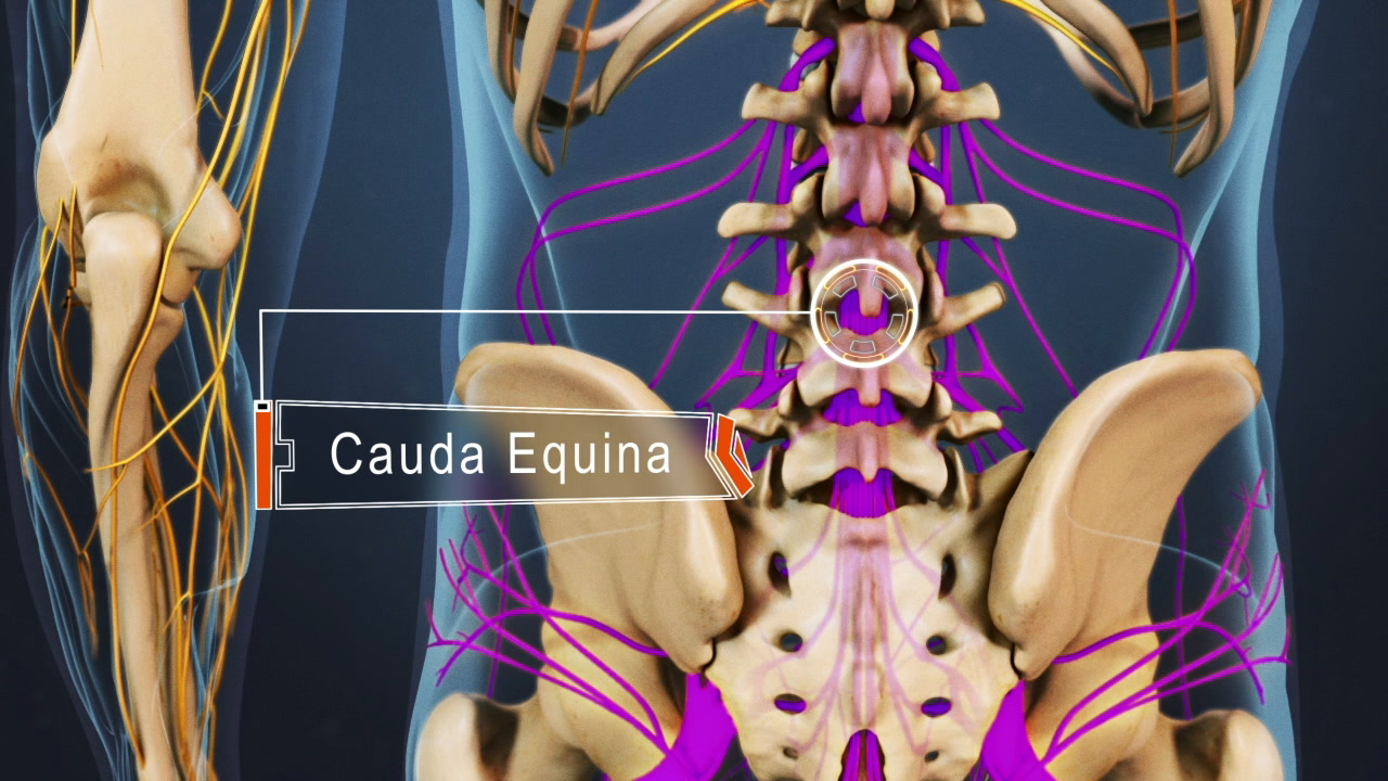
The lumbar spine includes the terminal end of the spinal cord and the cauda equina.
The spinal cord terminates in the lumbar spine. The exact point of termination varies among individuals; most commonly, it terminates at the level of the L1 or L2 vertebrae.
A few anatomical structures related to the lumbar spinal cord are highlighted below.
The terminal portion of the spinal cord in the lumbar region is cone-shaped and is called the conus medullaris.
The conus medullaris is made up of several neurons (nerve cells) and has 3 protective layers. Starting from the outermost layer, these are the dura mater, arachnoid mater, and pia mater. The pia mater tapers and continues down as the filum terminale at the end of the conus medullaris. 1 Nene Y, Jilani TN. Neuroanatomy, Conus Medullaris. [Updated 2019 Aug 3]. In: StatPearls [Internet]. Treasure Island (FL): StatPearls Publishing; 2019 Jan-. Available from: https://www.ncbi.nlm.nih.gov/books/NBK545227/
advertisementThe pia mater forms a delicate, fibrous strand of tissue called the filum terminale that extends down from the conus medullaris. This strand stabilizes the spinal cord by connecting the conus medullaris to the coccyx (end of the spine). 1 Nene Y, Jilani TN. Neuroanatomy, Conus Medullaris. [Updated 2019 Aug 3]. In: StatPearls [Internet]. Treasure Island (FL): StatPearls Publishing; 2019 Jan-. Available from: https://www.ncbi.nlm.nih.gov/books/NBK545227/
The dura mater and arachnoid mater are collectively called the dural sac or the thecal sac. This sac covers the conus medullaris, cauda equina, and each individual spinal nerve root as it courses out of the cauda equina and travels into the intervertebral foramen (bony opening through which the nerve exits the spinal canal). 2 Darby SA. General Anatomy of the Spinal Cord. In: Clinical Anatomy of the Spine, Spinal Cord, and Ans. Elsevier; 2014:65-97. doi:10.1016/b978-0-323-07954-9.00003-7
The space inside the arachnoid mater is filled with cerebrospinal fluid (CSF). Below the conus medullaris, this space is enlarged. This enlargement is called the lumbar cistern and contains CSF, the filum terminale, and the cauda equina. 2 Darby SA. General Anatomy of the Spinal Cord. In: Clinical Anatomy of the Spine, Spinal Cord, and Ans. Elsevier; 2014:65-97. doi:10.1016/b978-0-323-07954-9.00003-7

The cauda equina nerves travel down from the spinal cord.
A group of nerve roots that travel down from the spinal cord and the conus medullaris is called the cauda equina. The cauda equina contains nerve roots from L2 in the lumbar spine to Co1 in the coccygeal (tail bone end) spine. 1 Nene Y, Jilani TN. Neuroanatomy, Conus Medullaris. [Updated 2019 Aug 3]. In: StatPearls [Internet]. Treasure Island (FL): StatPearls Publishing; 2019 Jan-. Available from: https://www.ncbi.nlm.nih.gov/books/NBK545227/ Each nerve root from the cauda equina exits the spinal canal at its respective vertebral level, for example, the L4 nerve root exits between the L4 and L5 vertebrae.

Compression of the cauda equina, such as by a herniated disc, may cause severe pain and numbness in both legs.
The terminal tissues of the spinal cord in the lower back can get compressed, irritated, or abnormally stretched and typically cause serious medical problems. Possible conditions affecting this region along with the specific symptoms, signs, and causes are described below.
Compression of the conus medullaris in the lumbar region at L1-L2 (typically between T12 to L2) causes conus medullaris syndrome.
This syndrome is characterized by the following signs and symptoms 1 Nene Y, Jilani TN. Neuroanatomy, Conus Medullaris. [Updated 2019 Aug 3]. In: StatPearls [Internet]. Treasure Island (FL): StatPearls Publishing; 2019 Jan-. Available from: https://www.ncbi.nlm.nih.gov/books/NBK545227/ :
The most common causes of conus medullaris syndrome include spinal fracture, disc herniation, tumors, trauma, and collection of pus in the epidural space due to infection (epidural abscess). 1 Nene Y, Jilani TN. Neuroanatomy, Conus Medullaris. [Updated 2019 Aug 3]. In: StatPearls [Internet]. Treasure Island (FL): StatPearls Publishing; 2019 Jan-. Available from: https://www.ncbi.nlm.nih.gov/books/NBK545227/
Conus medullaris syndrome is a medical emergency and requires immediate treatment to preserve leg function.
An abnormal filum terminale can alter the stability of the conus medullaris and prevent its normal movement in the spinal canal. This condition is called tethered cord syndrome and causes the spinal cord to get abnormally stretched, especially during actions such as spinal flexion (bending forward). 1 Nene Y, Jilani TN. Neuroanatomy, Conus Medullaris. [Updated 2019 Aug 3]. In: StatPearls [Internet]. Treasure Island (FL): StatPearls Publishing; 2019 Jan-. Available from: https://www.ncbi.nlm.nih.gov/books/NBK545227/
Common signs and symptoms include 3 Tu A, Steinbok P. Occult tethered cord syndrome: a review. Child’s Nervous System. 2013;29(9):1635-1640. doi:10.1007/s00381-013-2129-1 :
While this condition is typically identified during periods of rapid growth in adolescence, the symptoms may not occur until adulthood. Common causes of tethered cord syndrome are split spinal cord (diastematomyelia), spinal cord lining problems (dermal sinuses), and lumps of fatty tissue in the spinal cord region (lipomas). 1 Nene Y, Jilani TN. Neuroanatomy, Conus Medullaris. [Updated 2019 Aug 3]. In: StatPearls [Internet]. Treasure Island (FL): StatPearls Publishing; 2019 Jan-. Available from: https://www.ncbi.nlm.nih.gov/books/NBK545227/
Compression of the cauda equina within the lumbar cistern can cause serious symptoms in the lower body.
The condition is called cauda equina syndrome and presents with the following signs and symptoms 4 Berg EJ, Ashurst JV. Anatomy, Back, Cauda Equina. [Updated 2018 Dec 6]. In: StatPearls [Internet]. Treasure Island (FL): StatPearls Publishing; 2019 Jan-. Available from: https://www.ncbi.nlm.nih.gov/books/NBK513251/ :
Cauda equina syndrome can present in 2 ways: acute onset, where the symptoms and signs occur rapidly, and insidious onset, where the condition begins as lower back pain and slowly progresses to bowel and urinary incontinence. 4 Berg EJ, Ashurst JV. Anatomy, Back, Cauda Equina. [Updated 2018 Dec 6]. In: StatPearls [Internet]. Treasure Island (FL): StatPearls Publishing; 2019 Jan-. Available from: https://www.ncbi.nlm.nih.gov/books/NBK513251/
advertisementCauda equina syndrome is most commonly caused by compression from a lumbar herniated disc. Compression may also occur due to tumors, cysts, stenosis (abnormal narrowing of the spinal canal), or trauma. Cauda equina syndrome is a medical emergency. Immediate treatment is crucial for the prevention of paralysis and preservation of leg function. 4 Berg EJ, Ashurst JV. Anatomy, Back, Cauda Equina. [Updated 2018 Dec 6]. In: StatPearls [Internet]. Treasure Island (FL): StatPearls Publishing; 2019 Jan-. Available from: https://www.ncbi.nlm.nih.gov/books/NBK513251/
If any of these spinal cord related problems are suspected, it is advisable to consult a doctor immediately. Spinal cord disorders may be treated nonsurgically or surgically depending on the cause and the overall health of the patient. The prognosis increases significantly when treated early.

Dr. Kara Beasley is a neurosurgeon practicing at Boulder Neurosurgical & Spine Associates, where she has several years of experience specializing in spine and brain surgery.